Desert Varish - Wet & Wild
UNM Scanning Electron Microscope Images by M. Spilde and P. Boston
(Click on an Image for a Larger Version)
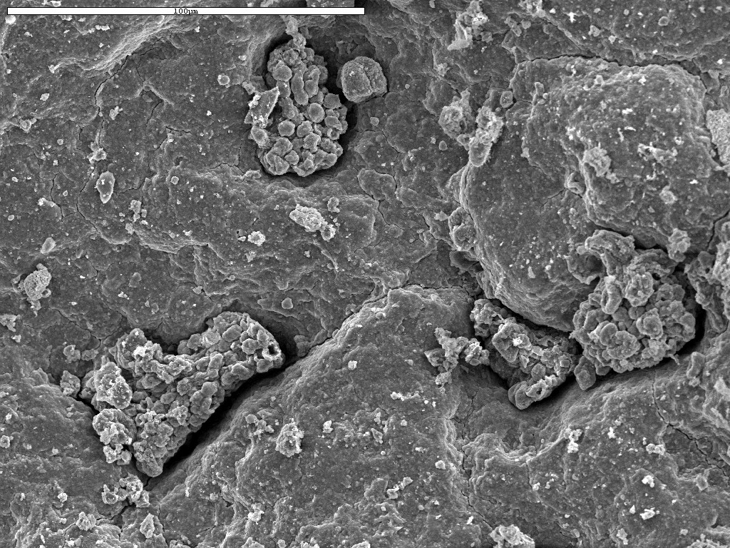
| These images are from two desert varnish pieces, soaked for 3 hours in water. They were then allowed to incubate for 40 hours, fixed in glutaraldehyde and then vacuum oven dried. Au/Pd coated. | |
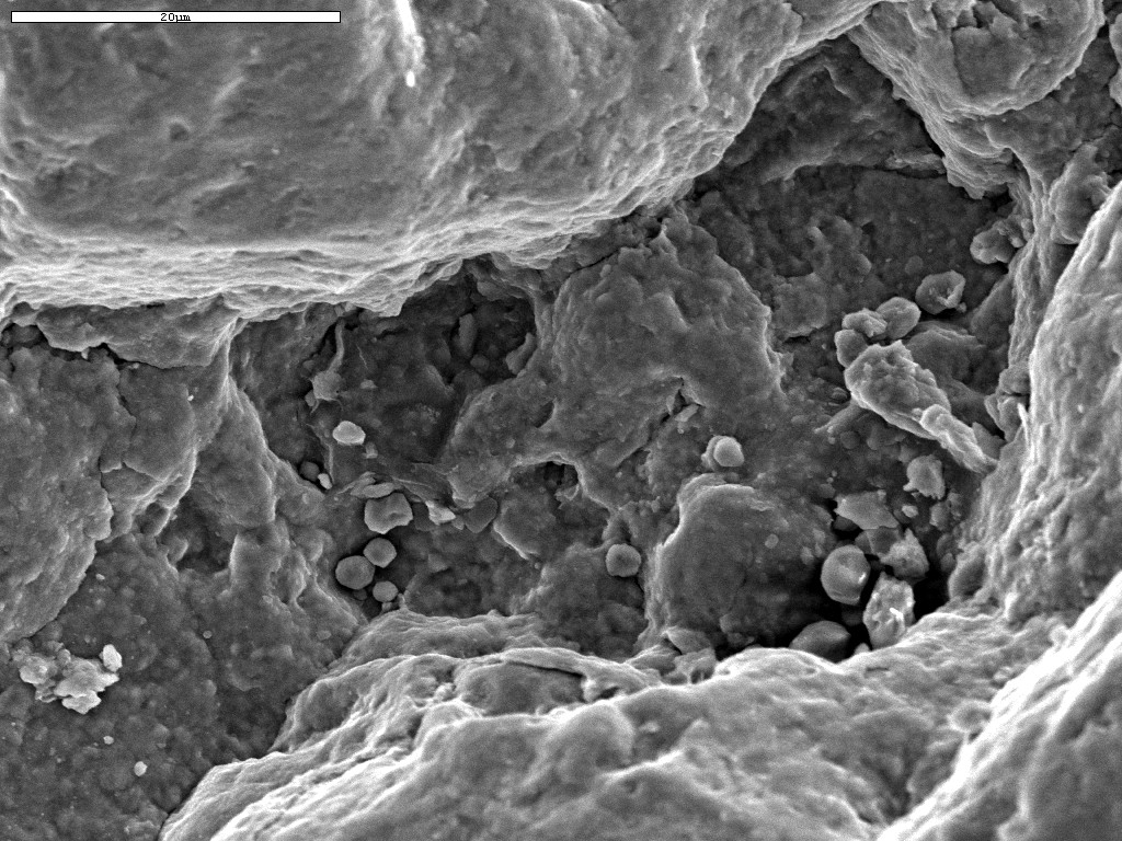
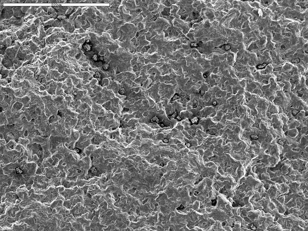 Whole field, note tiny colonies in many pits. |
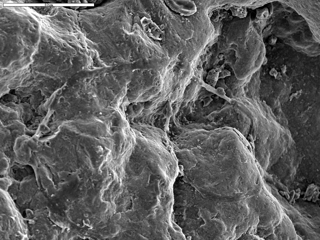 Closer detail of field. Very busy ... slime, live cells, preserved cells, a big mineral-impregnated filament, etc. |
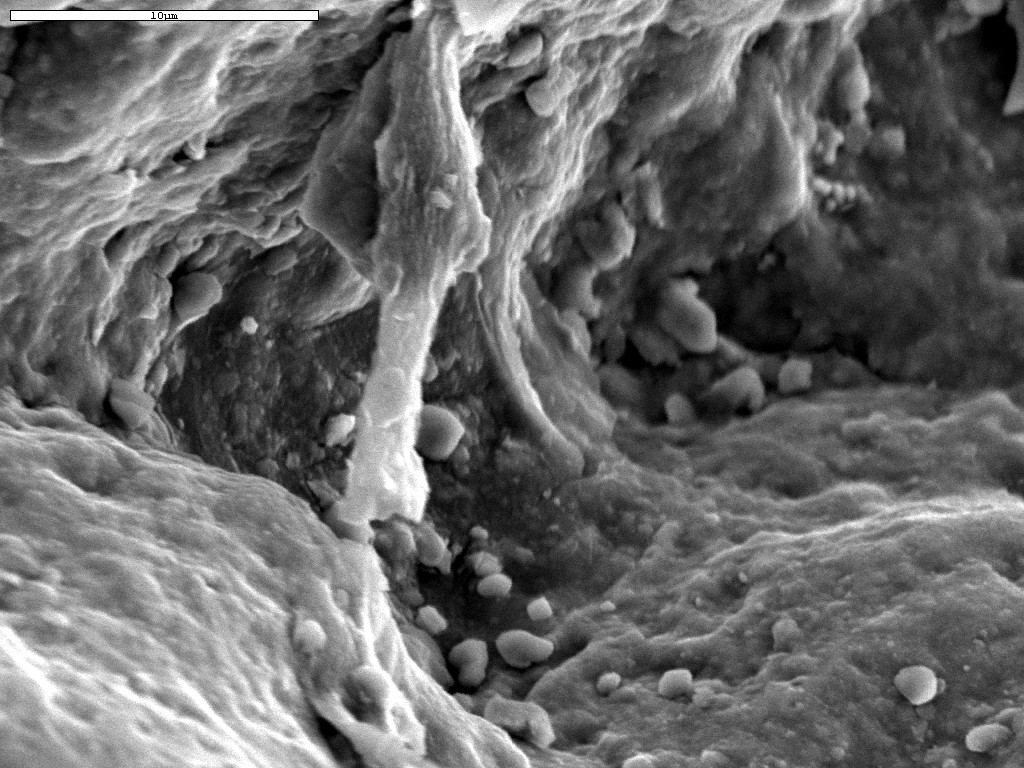 Juicy slime fibers, strong Mn signal in the EDS, lots of organic carbon. |
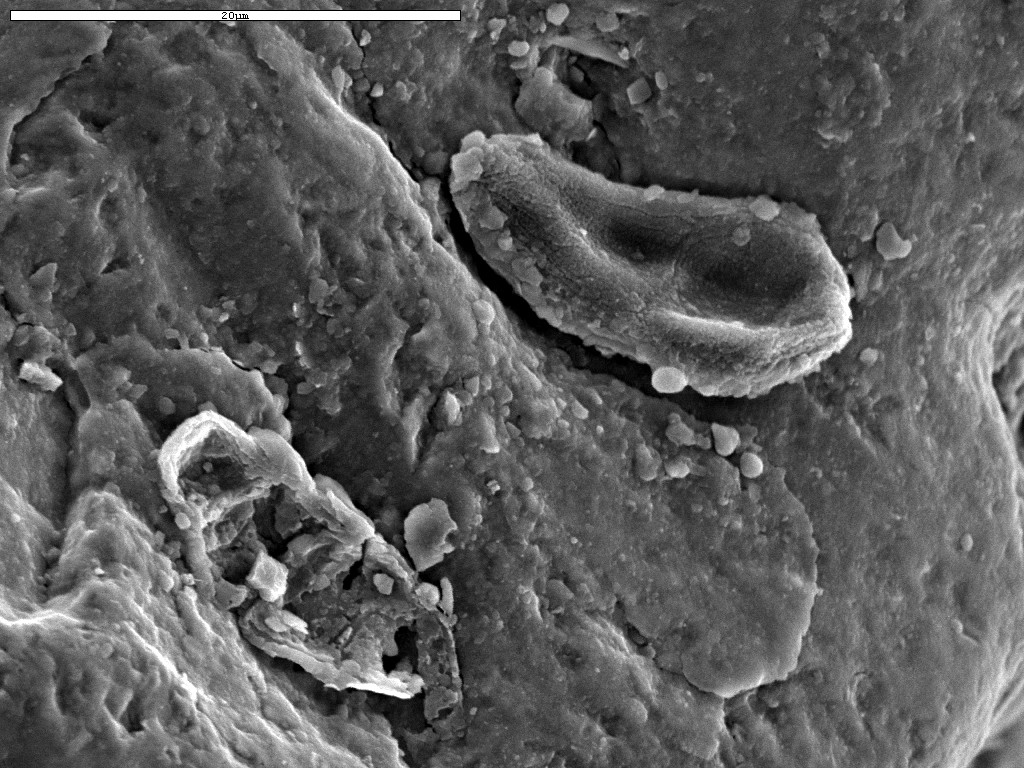 Upper right of field, juicy recently alive algal cell. Lower left of field, preserved cell remnants, perhaps of the same species as the live specimen. EDS shows lots of organic carbon in the live cell. The background appears to be silicate covered. The preserved cell also gives an exclusive silicate signal. |
 Iron oxide granules in pit. May or may not be coated organisms. |
 Juicy, organic carbon rich microcolony. Probably yeast or microcolonial fungi. Apparent budding off of small cells indicates a yeast. |
 Busy field, microcolony in pit, small single bacterial cell on left, filaments?, biogoo, and strong organic carbon and iron oxide EDS signals. |
 Close up of previous image. Left of field, approx. 1.6 micron juicy, carbonaceous bacterial cell. To the right of field, a more exciting find ... colony of small buried bacteria coated with biogoo and iron oxides. |
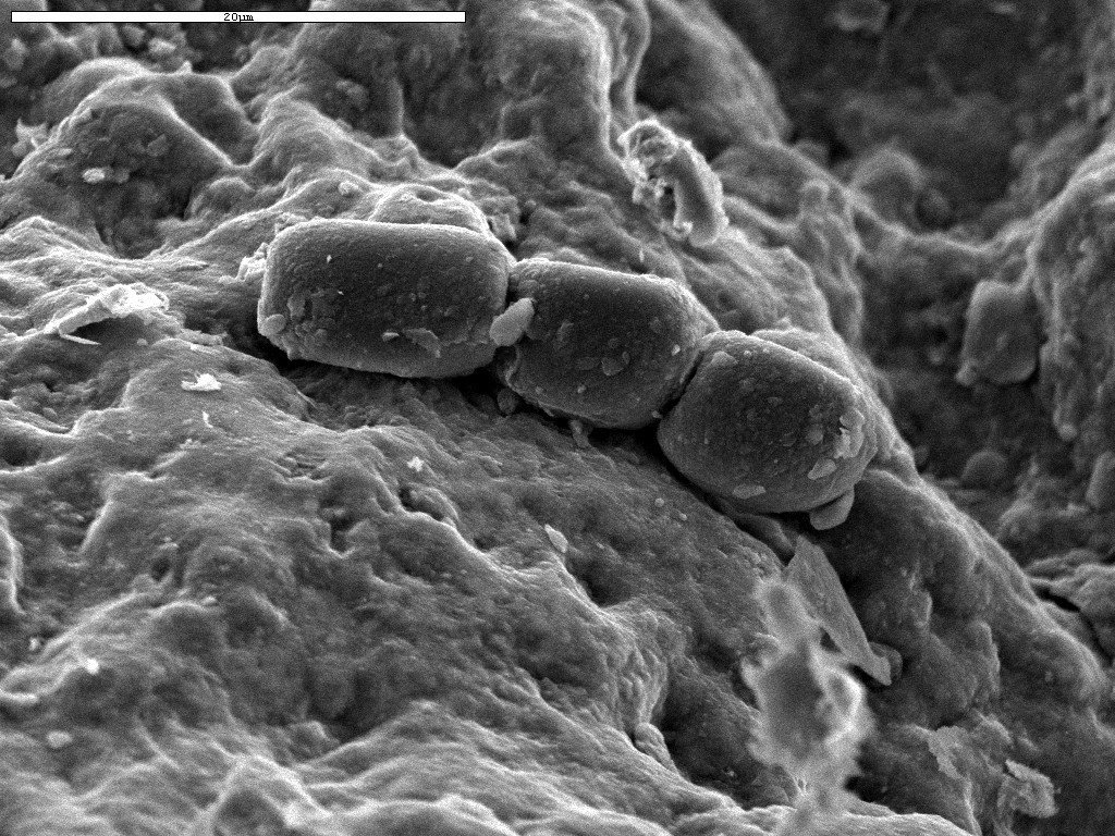 Large, rectangular algal cells. Small chain of a filamentous variety. |
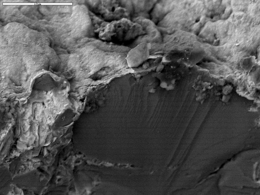 Broken edge of varnished piece. The Fe/Mn coating varies around 200-300 nm in thickness. |
The rest of the images are on a second piece of varnish, soaked for 3 hours in water, incubated for 40 hours, dried in vacuum oven (no glutaraldehyde step). | |
 These live cells are shriveled because of no glutaraldehyde fixation. |
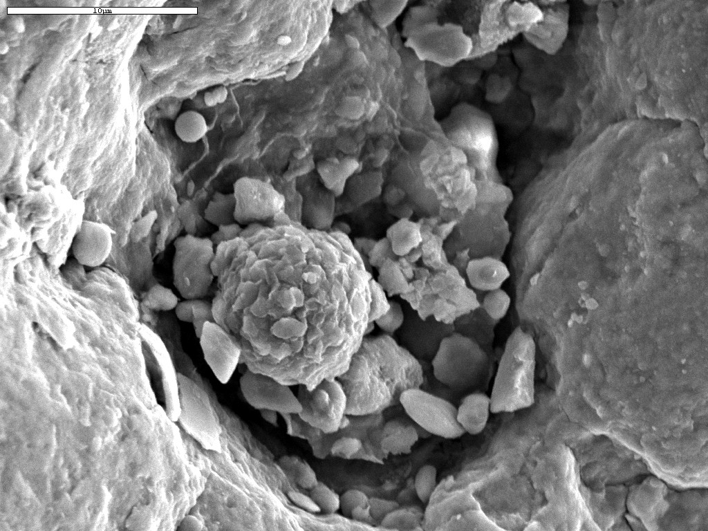
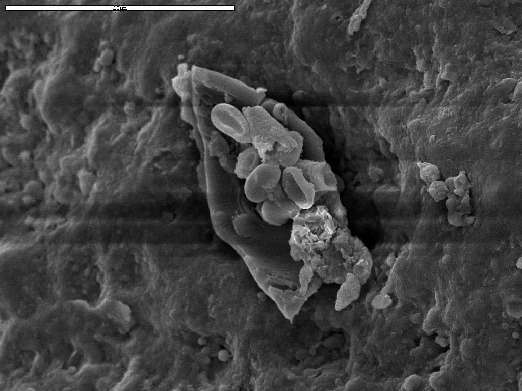
 Juicy, biogoo covered microcolony in pit. May be silicified but we couldn't get a decent EDS on this because it was in too deep a pit. Also on the right of field, a nice bicellular cyanobacterium. |
 Colonies in pits including the one in the previous image. Also, many different sized and shaped cells strewn around on the surface. Some appear mineralized, some degraded. |
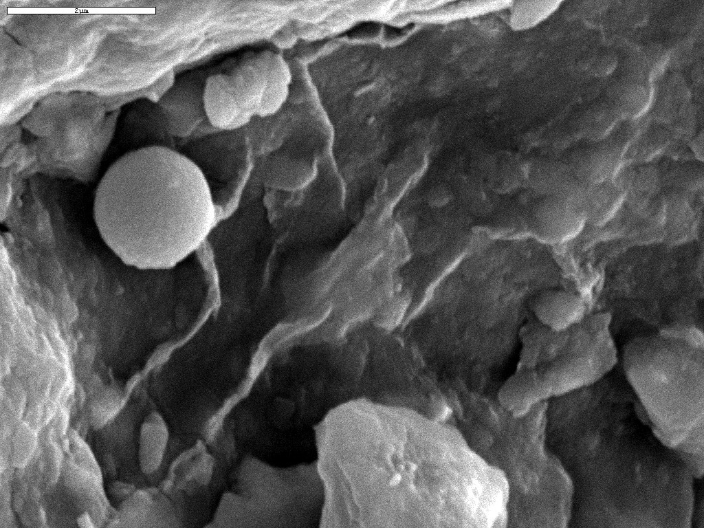
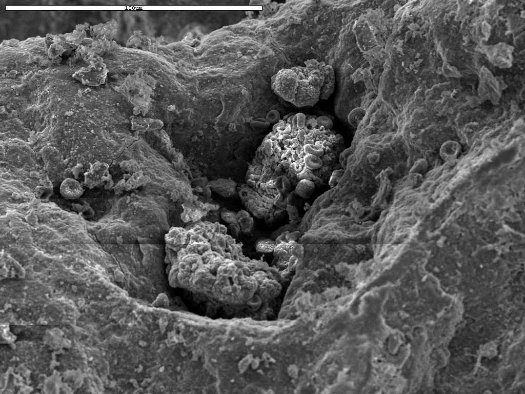 Juicy live algal and cyanobacterial cells (shriveled by drying without glut) strewn around the landscape. The microcolonies in the pits appear mineral impregnated. They may very well not be active but be preserved, although we could not get a clear EDS signal off the microcolony. |
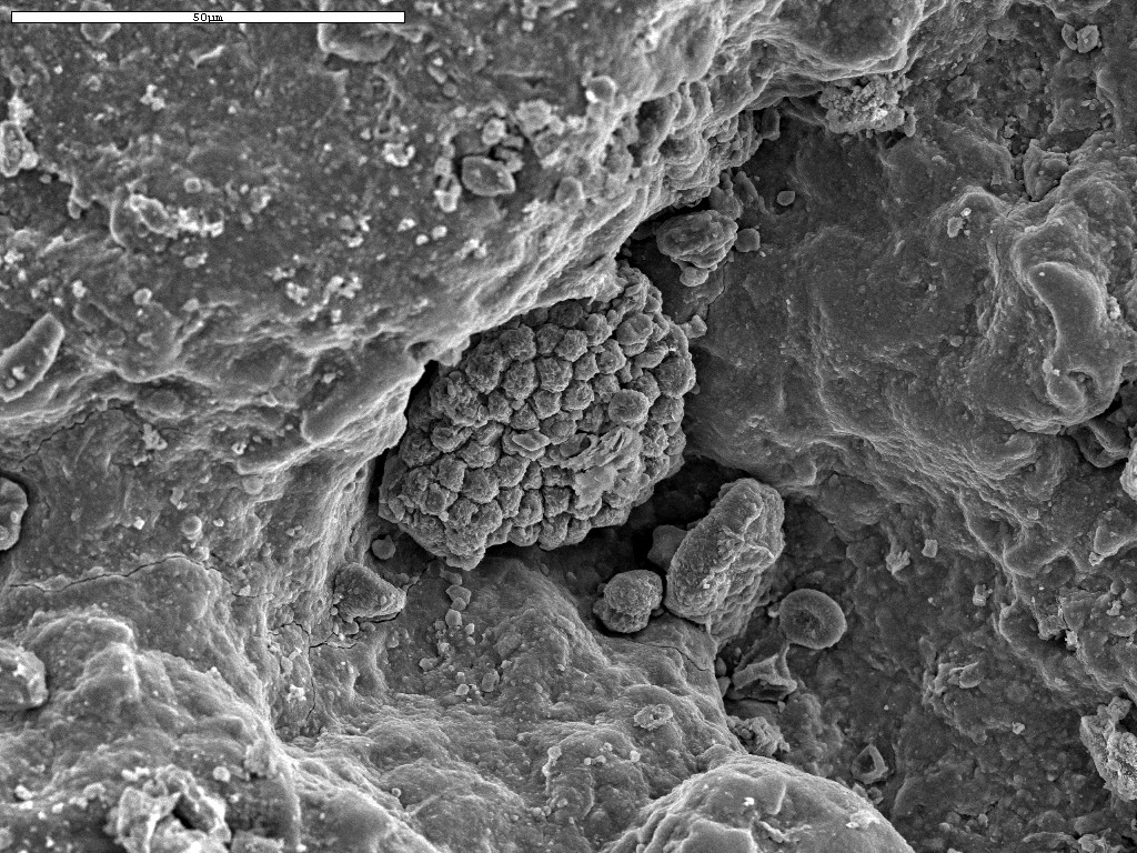 Microcolony in pit, many other cellular shapes strewn across the field of view. Couldn't get a good EDS in the pit, but did get a fairly clear indication of silicate coating. |
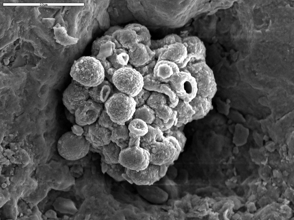
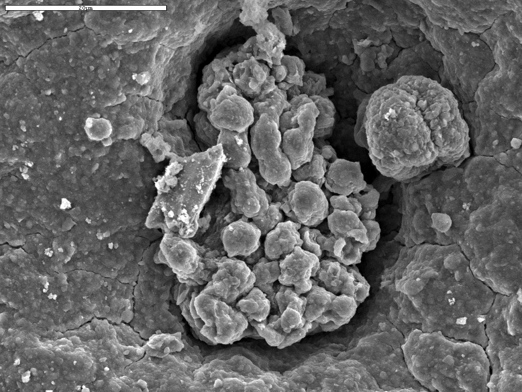
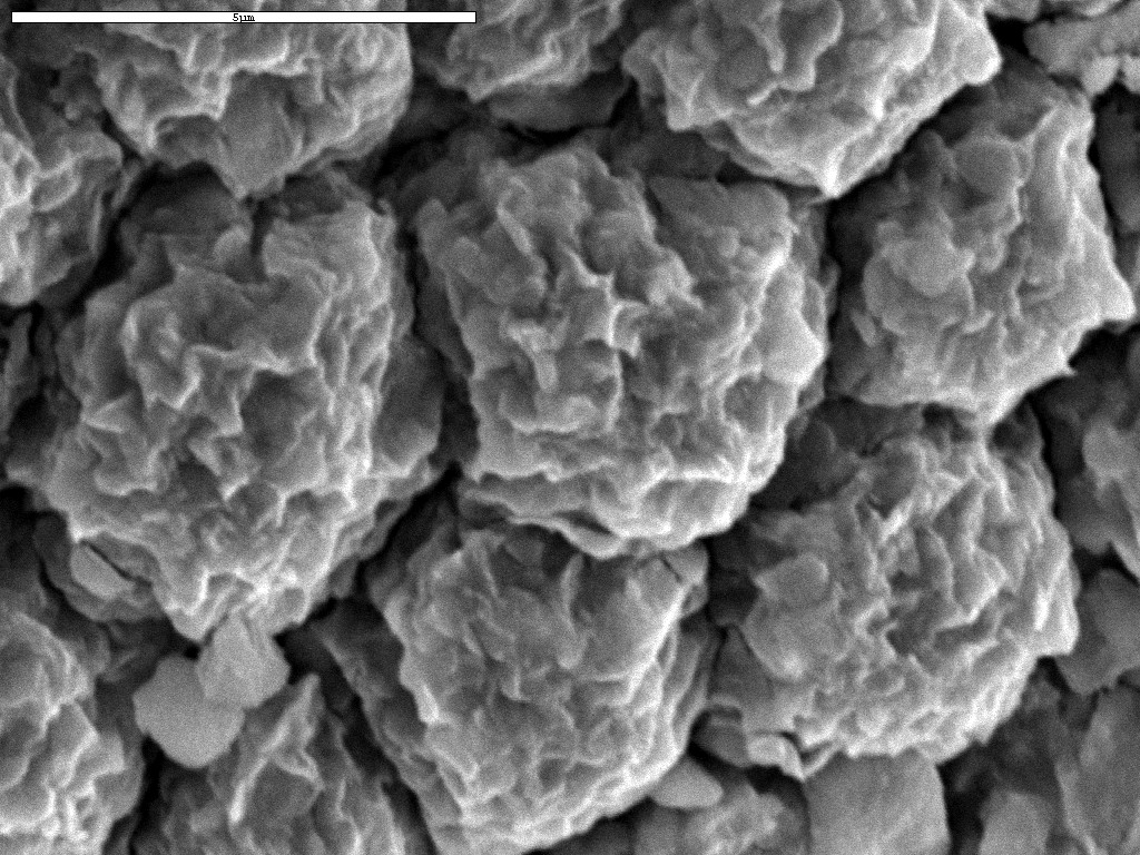 Closeup of microcolony in above image. The cells are about 4 microns in diameter. Their "wrinkly pea" appearance implies that they were very recently alive and growing before we dried them in the oven. |
Return to the Index Page